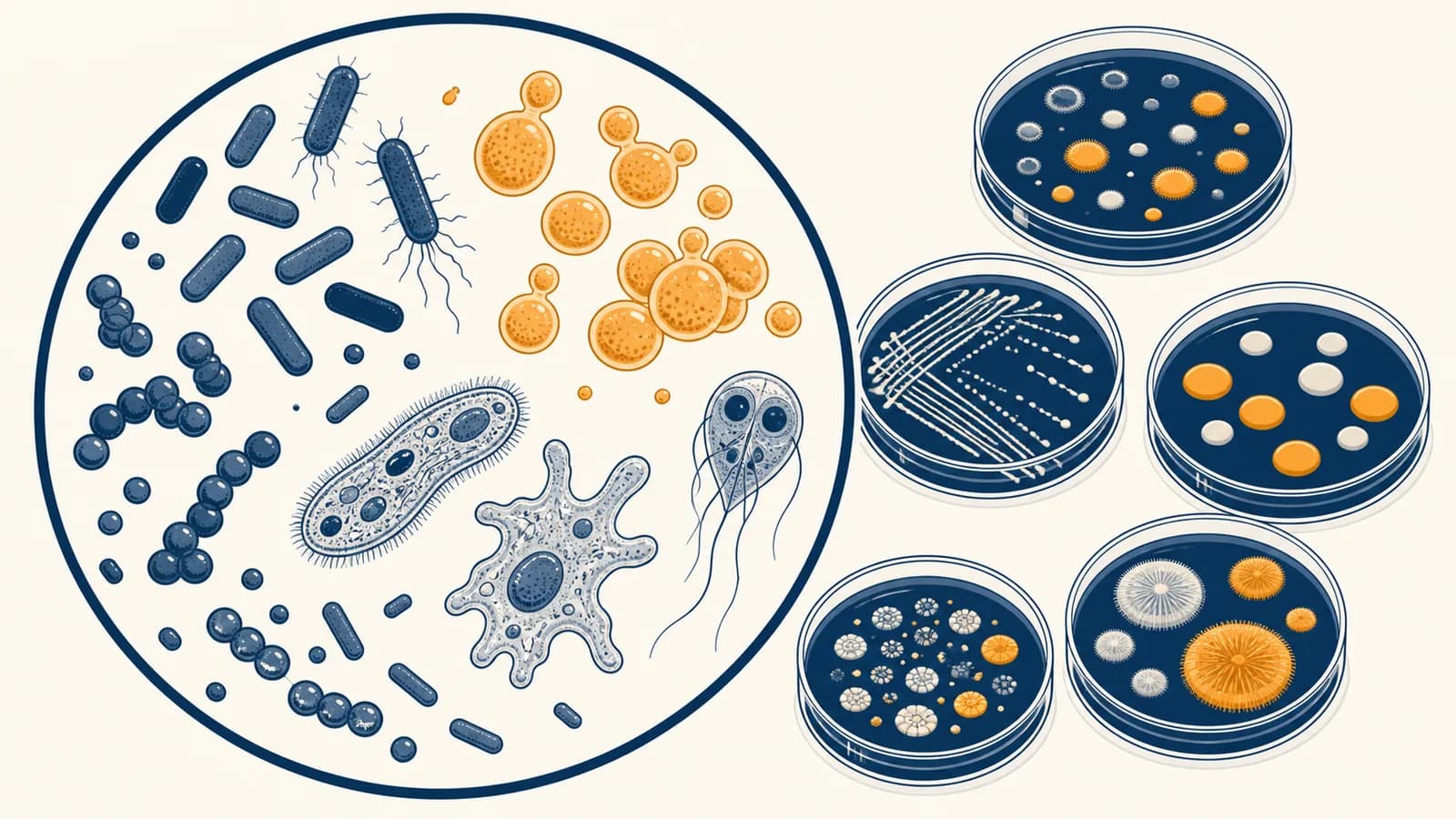
Microbiology is a branch of biology that studies microscopic organisms and acellular infectious agents, in the context of living systems, health, industry, and the environment. It explains what microorganisms are and how bacteria, archaea, fungi, protozoa, microscopic algae, and viruses reproduce, evolve, and affect other organisms. The subject exists because things too small to see directly can still cause disease, make food, recycle matter, alter climate, and control much of the chemistry around us. Microbiology connects cell structure to visible consequences, such as bread rising, a wound becoming infected, or sewage becoming safe enough to release.
What microbiology actually is
Microbiology is the study of organisms and biological agents that usually require magnification, culture methods, chemical tests, or genetic tools to detect and identify. It asks what they are made of, how they obtain energy, and how they interact with hosts and habitats.
The word microbe is a practical umbrella term, not one branch on the tree of life. Bacteria and archaea are cellular organisms without a nucleus. Fungi and protozoa are eukaryotes whose cells contain a nucleus. Microscopic algae are photosynthetic eukaryotes. Viruses are packages of genetic material that reproduce only by using a host cell. These groups differ so much that a method that kills a bacterium may do nothing to a virus or fungal spore.
A microbiologist rarely identifies an unknown sample by appearance alone. Cell shape gives clues, but many unrelated organisms look similar under a light microscope. Identification usually combines evidence: staining behavior, growth conditions, chemical reactions, immune recognition, and DNA or RNA sequences. Each test narrows the possibilities.
This is one reason microbiology sits inside the wider study of Biology. It uses the same principles as genetics, evolution, chemistry, and physiology, but applies them to small systems that can multiply quickly and be tested in controlled conditions.
How microbial cells are built and classified
Microbial cells are classified by ancestry and by observable features such as cell walls, membranes, metabolism, shape, and genetic sequence. Their structures solve the same basic problems: keeping an inside separate, copying information, making proteins, obtaining energy, and responding to conditions.
Bacteria and archaea organize life without a nucleus
A bacterial cell usually has a plasma membrane, cytoplasm, ribosomes, and one main circular chromosome in a region called the nucleoid. Many bacteria also carry plasmids, which are smaller DNA molecules that copy separately. A cell wall outside the membrane resists osmotic pressure. Some species add a capsule, flagella for movement, or pili that help with attachment and DNA transfer.
Archaea have the same compact plan, but they are not unusual bacteria. Their membrane lipids have different chemical linkages, their cell walls lack bacterial peptidoglycan, and parts of their information processing machinery resemble those of eukaryotes. Genetic evidence places bacteria, archaea, and eukaryotes in three distinct domains.
Small does not mean simple. A bacterium can sense nutrients, regulate thousands of genes, repair DNA, move toward favorable chemicals, and coordinate behavior with neighboring cells.
Microbial eukaryotes contain specialized compartments
Yeasts are single celled fungi, while molds grow as networks of filaments called hyphae. Protozoa are a diverse collection of mostly single celled eukaryotes that often move or consume other cells. Microscopic algae capture light energy in chloroplasts. Their nuclei separate DNA from the cytoplasm, and organelles divide cellular work among compartments.
Classification now follows evolutionary relationships
Older classification relied heavily on shape and metabolism. Modern classification compares inherited sequences, especially genes found across many organisms. Similar sequences suggest shared ancestry. The result is a branching family tree, not a ladder from simple to advanced. A round cell and another round cell may be distant relatives that independently kept the same useful shape.
The structures named here are developed more fully in cell membranes, organelles, and division. Microbiology adds the practical question: how can those features reveal what is present in an unknown culture?
How microbial growth works
Microbial growth usually means an increase in cell number, not an increase in one cell's size. Cells take in material, duplicate their contents, and divide. Population growth depends on nutrients, temperature, water, acidity, oxygen, waste, and competition.
Many bacteria reproduce by binary fission. The chromosome is copied, the two DNA copies move apart as the cell lengthens, and a division wall forms between them. One cell becomes two descendants. Budding yeast grows a smaller outgrowth, copies its nucleus, and separates the bud. Fungal hyphae extend at their tips and branch through a food source.
Transport proteins bring in nutrients, while sensors detect temperature, acidity, oxygen, and chemical signals.
Enzymes break down nutrients, generate ATP, and build membrane lipids, proteins, RNA, and DNA.
DNA replication makes the inherited instructions needed by each descendant cell.
Cell components are distributed and a new boundary separates the descendants.
Under steady ideal conditions, repeated division produces exponential growth. If every cell divides once per generation and none die, the population doubles each generation.
If 100 cells divide every 30 minutes for 2 hours, then and cells.
Real cultures do not grow exponentially forever. In a closed flask, cells first adjust to the medium during lag phase. Rapid division follows in exponential phase. Nutrient shortage and waste accumulation then make cell production roughly balance cell death in stationary phase. If harmful conditions continue, viable cell numbers decline. A culture can therefore look cloudy even after many cells have died, because dead cells and fragments still scatter light.
Viruses versus living cells
Cells carry out metabolism, maintain internal conditions, and reproduce using their own cellular machinery, while viruses must enter suitable host cells to make new virus particles. A virus is biological and can evolve, but it is not an independently living cell.
Contains membrane, cytoplasm, ribosomes, and DNA. It can take in nutrients, generate ATP, make proteins, and divide when conditions permit.
Contains DNA or RNA inside a protein coat, sometimes wrapped in a host derived membrane. Outside a host, it does not make ATP, grow, or manufacture proteins.
Viral replication begins with recognition. Proteins on the virus bind particular molecules on a host cell, which helps determine which species and tissues the virus can infect. The viral genome then enters the cell or is released inside it. Host ribosomes translate viral instructions, and host or viral enzymes copy the genome. Newly made genomes and proteins assemble, then leave by bursting the cell or budding through its membrane.
Antibiotics target bacterial features such as cell wall synthesis or bacterial ribosomes. Viruses lack those targets, so antibiotics do not treat viral infections. Antiviral drugs instead interfere with a virus specific step, such as genome copying, protein processing, or entry. Because host and virus processes are intertwined, finding selective viral targets can be difficult.
How microbes obtain energy and matter
Microbes survive by coupling energy releasing reactions to cell work and by obtaining carbon for cellular material. They may use light or chemicals for energy, and carbon dioxide or organic molecules for carbon, producing combinations of metabolism found across many habitats.
Respiration transfers electrons from an electron donor through carriers to a final electron acceptor. The transfer pumps protons across a membrane. Protons flow back through ATP synthase, which uses that flow to make ATP. In aerobic respiration, oxygen is the final electron acceptor. Some microbes instead use substances such as nitrate or sulfate when oxygen is absent.
Fermentation does not use an external final electron acceptor or a respiratory electron transport chain. It rearranges organic molecules so that electron carrying molecules can be recycled, allowing glycolysis to continue making a smaller supply of ATP. Yeast fermentation converts sugars into ethanol and carbon dioxide. Lactic acid bacteria convert sugars mainly into lactic acid, which lowers pH and changes food texture and flavor.
| Metabolic strategy | Energy source | Carbon source | Familiar setting |
|---|---|---|---|
| Photoautotrophy | Light | Carbon dioxide | Cyanobacteria in illuminated water |
| Chemoautotrophy | Inorganic chemical reactions | Carbon dioxide | Nitrifying microbes in soil and filters |
| Chemoheterotrophy | Organic chemical reactions | Organic molecules | Yeast in dough and many gut bacteria |
Metabolic diversity lets microbial communities pass materials along. One species releases a product that another consumes. In oxygen free sediment, for example, fermenters break complex molecules into smaller compounds, and other microbes use those compounds in respiration or methane production. The community performs a chemical sequence that no member completes alone.
How microbes exchange genes and evolve
Microbial populations evolve when mutation and gene transfer create inherited variation, then environmental conditions change which variants leave more descendants. Bacteria can also acquire genes from nearby cells or free DNA, so useful traits may spread without parent to offspring reproduction.
Mutation creates new sequence variants
DNA copying is accurate but not perfect. A change may have no effect, harm the cell, or alter a useful trait. Mutations do not appear because a cell needs them. Instead, conditions select among variants already present or newly produced. If an antibiotic kills susceptible cells while a resistant mutant survives, the next population contains a greater fraction of the resistant lineage.
Horizontal gene transfer moves existing instructions
Transformation occurs when a cell takes up free DNA from its surroundings. Transduction occurs when a bacterial virus carries DNA between cells. Conjugation involves direct contact and commonly transfers a plasmid through a cell to cell connection. These routes can move genes for toxin production, new metabolic abilities, or drug resistance.
A patient has a bloodstream infection. The laboratory grows the bacterium and exposes it to several antibiotics. Clear zones around some antibiotic sources show inhibited growth, while growth beside another suggests resistance. The result helps the clinical team choose a drug, but the patient's condition, infection site, allergies, and drug behavior in the body also matter.
Selection also occurs outside hospitals. Antibiotics used in people, animals, or laboratory cultures create a strong filter wherever active drug reaches microbes. Hygiene and correct prescribing reduce opportunities for resistant strains to spread. The immune processes that recognize and remove microbes belong to the biology of immune defense, which works alongside antimicrobial treatment rather than replacing it.
How microbiology shows up in human health
Medical microbiology identifies infectious agents, explains how they spread and cause damage, and supports prevention and treatment. Disease depends on the microbe, its route into the body, the dose, the infected tissue, and the host's defenses and health.
A pathogen must complete several tasks. It reaches a host, enters through a suitable route, attaches or gains access to tissue, obtains resources, avoids removal long enough to multiply, and exits or reaches another host. Virulence factors are microbial features that help with these tasks. A capsule may hinder engulfment by immune cells. An adhesion protein may bind a tissue surface. A toxin may damage cells or disrupt signaling.
Symptoms can come from direct microbial damage, toxins, or the body's response. Fever and inflammation are host responses. Diarrhea may result from a toxin changing ion movement across intestinal cells, which draws water into the gut. Tissue damage during an infection can therefore reflect both microbial action and defense mechanisms.
Colonization is not the same as infection. A microbe can live on a body surface without invading tissue or causing damage. Infection means it has entered and multiplied in a host, while infectious disease means that process has impaired normal function.
Diagnosis starts with a sample taken from the relevant site, such as blood, urine, sputum, stool, skin, or cerebrospinal fluid. Microscopy can reveal cells, fungi, or parasites. Culture can isolate living organisms and test their drug response. Antigen tests detect microbial molecules. Nucleic acid tests amplify and detect selected DNA or RNA sequences. A result must be interpreted in context because contamination, harmless colonization, or traces of a past infection can mislead.
How microbiology shows up in food, industry, and ecosystems
Applied microbiology uses microbial metabolism to make products, prevent spoilage, treat waste, and track environmental processes. The same growth rules that explain infection also govern yogurt cultures, brewing yeast, sewage reactors, compost, soil fertility, and contamination control in factories.
Fermentation turns microbial metabolism into food production
Bakers mix yeast into dough, where it consumes available sugars and releases carbon dioxide. Gas becomes trapped in the elastic dough network, expanding it. During yogurt production, bacteria convert lactose into lactic acid. Falling pH changes milk proteins so they form the characteristic gel and makes conditions less suitable for many spoilage organisms.
Bioreactors make cells work under controlled conditions
A bioreactor holds cells or enzymes while operators control mixing, temperature, acidity, nutrients, and gas supply. Microbes can produce enzymes, vitamins, organic acids, medicines, and proteins encoded by inserted genes. Production only succeeds if the chosen strain stays healthy and contaminants are excluded. A contaminating microbe may consume feed, alter the product, or introduce a safety hazard.
Microbes run elemental cycles
Decomposers digest dead material and release smaller molecules that other organisms can use. Nitrogen fixing microbes convert atmospheric nitrogen gas into ammonia, a form that can enter biological molecules. Nitrifying microbes oxidize ammonia into nitrite and then nitrate. Denitrifying microbes can return nitrogen to the atmosphere under low oxygen conditions. These reactions link microbial enzymes to soil fertility and water quality.
Those exchanges connect microbiology to organisms, communities, and nutrient cycles. A forest, lake, farm, or human intestine cannot be explained by listing large organisms alone. Microbial reactions often control which chemical forms are available to the rest of the system.
Robert Hooke's Micrographia records microscopic structures and gives cells their name after observations of cork.
Antonie van Leeuwenhoek describes tiny living organisms seen with his single lens microscopes.
Louis Pasteur's swan neck flask experiments show that microbial growth in broth depends on contamination by particles from the environment.
Alexander Fleming notices that a mold contaminant inhibits nearby staphylococcal bacteria, an observation that contributes to later penicillin development.
These experiments did more than add names to a timeline. They established methods that still define the subject: observe with instruments, separate possible causes, grow organisms under controlled conditions, and connect a particular biological agent to a repeatable effect.
What the microbiome actually means
A microbiome is the microbial community in a defined habitat together with its genes, activities, and surrounding conditions. A gut microbiome, soil microbiome, and skin microbiome are different systems, and no single list of microbes defines health in every person or place.
Microbiota often means the organisms themselves, while microbiome is frequently used more broadly. Researchers study these communities by culturing members, sequencing marker genes, sequencing all sample DNA, measuring RNA, or analyzing metabolites. Each method answers a different question. DNA can show that genetic material is present, but it does not by itself prove that an organism is alive or a gene is active.
Members of a microbiome compete for space and nutrients, alter acidity and oxygen, exchange metabolites, and interact with host tissues. In the intestine, microbial enzymes can digest compounds that human enzymes do not break down. The resulting products may feed other microbes or be absorbed by the host. Effects depend on amounts and context, so dividing every species into simply good or bad gives a poor model.
A detected microbe caused the person's symptom, or one supplement will create a universally healthy community.
Detection establishes presence. Causation needs controls, timing, mechanism, and often experiments. Useful interventions depend on the organism, dose, host, diet, and medical setting.
How cleaning, disinfection, and sterilization differ
Cleaning removes dirt and many microbes, disinfection inactivates many disease causing microbes on objects, and sterilization eliminates all viable microorganisms, including resistant bacterial spores. The correct method depends on the material, the likely agent, and the risk if any survive.
Soap helps lift oils and particles from skin so water can carry them away. Friction and rinsing contribute to removal. Some disinfectants damage membranes, denature proteins, or oxidize cellular components. Their performance depends on concentration, contact time, temperature, surface coverage, and interfering material. Applying a product and wiping it away immediately may not allow the labeled contact time.
Heat transfers energy into cells and can disrupt membranes and proteins. An autoclave uses saturated steam under pressure so it can reach temperatures above the normal boiling point of water and contact suitable surfaces. Pressure itself is not the main killing agent. The hot steam is. Items must be arranged so steam reaches them, and the process must be validated rather than assumed from a warm chamber.
A cutting board used for raw poultry carries food residue and may carry pathogens. First remove residue with detergent and water. Then use a food surface method intended for sanitation, following its directions. Spraying disinfectant onto visible grease is less reliable because the material can shield microbes and consume the active chemical.
Pasteurization is different from sterilization. It applies a controlled treatment that reduces pathogens and spoilage organisms while limiting damage to the food. Some organisms or spores may remain, which is why pasteurized products can still require refrigeration and have finite shelf lives.
How scientists know which microbe caused an effect
Microbiologists connect a microbe to an effect by combining detection, isolation, controlled comparison, mechanism, and repeatability. Strong evidence shows more than correlation: the suspected agent appears in the relevant setting and changing its presence or activity predictably changes the outcome.
Classical disease studies used ideas associated with Koch's postulates: find the organism in diseased hosts, isolate it, reproduce disease in a suitable host, and recover the same organism. The framework was powerful, but biology supplies exceptions. Some pathogens cannot be grown in ordinary culture. Some cause disease only in humans. Some infected people carry an agent without symptoms. Ethical limits prevent deliberate human infection in many cases.
Modern evidence can connect a specific gene product to a mechanism. Researchers might compare a normal strain with a mutant missing a suspected toxin gene. If the mutant loses the damaging effect, then restoring the gene restores it, the gene has a stronger causal case. Microscopy may show where the microbe sits, chemical assays may detect the toxin, and host cell experiments may reveal the disrupted pathway.
Contamination controls matter at every stage. A blank sample that goes through the same tubes, reagents, and machines can reveal DNA or cells introduced by the procedure. Positive controls show that the test can detect its target. Without controls, an impressive signal may describe the equipment rather than the original sample.
Four mistakes people make with microbes
Common errors come from treating all microbes as harmful, confusing detection with disease, assuming products work instantly, and imagining evolution as a deliberate response. Correcting these errors makes health claims, laboratory results, food advice, and environmental news much easier to judge.
1. Treating every microbe as a germ
Germ usually means a disease causing agent, while microbe covers organisms with many roles. Most microbial encounters do not produce disease. Some microbes are harmless passengers, some compete with pathogens, and others perform fermentation or nutrient cycling. Safety depends on identity, amount, location, and host condition.
2. Reading a positive test as a complete diagnosis
A positive result means the test detected its target under its stated rules. It may not show that the target is alive, causing symptoms, or present in the affected tissue. Clinicians interpret test performance alongside symptoms, sampling time, exposure, and other possible causes.
3. Assuming more disinfectant always works better
Disinfectants are tested at defined concentrations and contact times. Using too little may fail, but mixing products or increasing concentration can create toxic fumes, damage surfaces, or add risk without improving the result. The product label defines the intended organism, surface, dilution, and exposure.
4. Saying microbes mutate to escape a threat
Mutation generates variation without planning. Antibiotic exposure changes survival, so variants with resistance leave more descendants. The population changes even though no cell predicted the drug. This distinction separates the origin of variation from natural selection acting on it.
Microbiology makes the rest of biology visible in action
Microbiology turns broad biological principles into processes that can be observed, measured, and tested: membranes control exchange, enzymes drive metabolism, genes carry information, populations evolve, and communities reshape environments. Small scale does not reduce importance; it makes cause and effect easier to isolate.
A useful habit is to trace any microbial claim through four questions. What organism or agent is present? What evidence identifies it? What mechanism could produce the effect? What comparison rules out another cause? Those questions work for an infection report, a probiotic advertisement, a food recall, or a claim about soil health.
Look for microbial processes in ordinary places. Bread dough records carbon dioxide production. Compost heat records rapid metabolism. A refrigerated leftover slows growth by lowering reaction rates, but does not reset the food to a sterile state. A cloudy pond may reflect photosynthetic microbes responding to nutrients and light. Each observation joins cell scale events to the larger living system.
The takeaway: Microbiology explains how microscopic cells and viruses are built, reproduce, exchange matter and information, and change hosts and environments. Follow the mechanism, then check the evidence that links the microbe to the outcome.