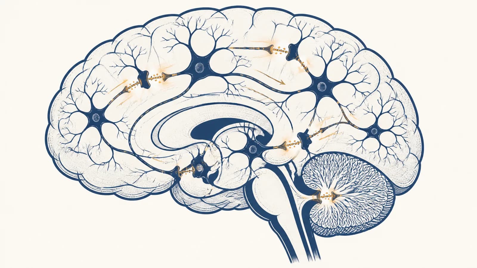
Neuroscience is a branch of biology that explains how nervous systems generate sensation, movement, thought, emotion, and behavior, in the context of living organisms. It studies how the brain works, how neurons communicate, what the spinal cord and nerves do, and how experience changes neural circuits. The field exists because behavior cannot be explained fully by naming body parts: scientists need to connect events inside cells to actions by a whole animal. Neuroscience therefore works across several scales, from ion channels in a membrane to decisions made by networks containing many cells.
What neuroscience actually is
Neuroscience is the study of nervous systems at several connected levels: molecules control cells, cells form circuits, circuits produce behavior, and behavior changes circuits. Its central task is to explain those links with evidence from anatomy, physiology, experiments, computation, and clinical cases.
The nervous system receives information, estimates what it means, selects responses, and adjusts future responses. Those jobs are distributed. Receptors in the eye convert light into electrical signals. Networks in the brain compare the signals with stored information. Motor pathways send commands toward muscles. Chemical changes at synapses can make a later response different from the first one.
This pipeline is useful, but it is not a row of isolated boxes. Signals circulate through feedback loops. Movement changes the sensory input arriving from muscles and joints. Attention changes which sensory signals have more influence. A result can become a new cause milliseconds later.
Neuroscience belongs inside the wider study of Biology because a brain is living tissue. It uses energy, regulates genes, builds proteins, responds to hormones, repairs some damage, and fails when its cells or blood supply are disrupted. Psychology describes patterns in thought and behavior. Neuroscience asks what biological processes can produce those patterns.
What neurons and glia actually are
Neurons are excitable cells specialized to receive, combine, and send information, while glia are diverse cells that support, insulate, defend, nourish, and regulate neural tissue. Both are active parts of nervous system function, and neither works properly without the other.
A neuron has regions built for information flow
A typical neuron has dendrites that receive many inputs, a cell body that maintains the cell, an axon that carries output, and axon terminals that communicate with target cells. The exact shape varies. A spinal motor neuron may extend an axon a long distance to muscle, while a local interneuron may connect only nearby cells.
The cell membrane separates fluids with different ion concentrations. Protein pumps maintain those differences, and protein channels let selected ions cross. This arrangement stores potential energy, rather like separated electric charge in a battery. The comparison has limits because ions also move by diffusion and channels change state, but it captures why the membrane can power rapid signals.
- Dendrites receive many synaptic inputs.
- The cell body maintains the cell and combines influences.
- The axon conducts an output signal.
- Axon terminals signal to other cells.
The basic cell machinery comes into clearer view through cell membranes, organelles, and transport. Neurons contain nuclei, ribosomes, mitochondria, and a cytoskeleton like other animal cells. Their unusual proportions create unusual logistical problems. Materials made near the nucleus must be transported along axons, sometimes to terminals far from the cell body.
Glia do more than hold neurons in place
Astrocytes help regulate the chemical environment around synapses and interact with blood vessels. Oligodendrocytes in the central nervous system and Schwann cells in peripheral nerves wrap axons with myelin. Microglia are immune cells of the central nervous system that respond to injury and help reshape connections. Other glial types line fluid spaces or support neurons in peripheral ganglia.
Myelin changes signal timing. It electrically insulates sections of an axon, so an action potential is regenerated mainly at gaps called nodes of Ranvier instead of continuously along every patch of membrane.
Faster is not always the only goal. Precise arrival time matters because a neuron may combine inputs that reach it within a narrow interval. Changes in myelin can therefore alter coordination within a circuit as well as conduction speed.
How neurons generate and transmit signals
Neurons signal by controlling ion flow across their membranes. Inputs shift membrane voltage, and if the axon reaches threshold, voltage gated channels produce an action potential that travels to terminals, where it can trigger chemical communication with the next cell.
A resting membrane stores usable energy
A resting neuron has an electrical potential across its membrane. Selective permeability and ion concentration differences create this voltage. The sodium potassium pump supports the gradients by using ATP to move three sodium ions out for every two potassium ions moved in. It does not single handedly create each action potential. Instead, it maintains the conditions that make repeated signaling possible.
The unequal exchange moves one net positive charge out of the cell during each cycle. More importantly, the pump preserves the sodium and potassium gradients that other channels use. Blocking the pump does not erase the membrane voltage instantly, but the gradients eventually run down.
If membrane voltage differs from an ion's equilibrium potential, open channels permit a current whose size also depends on conductance.
This relation explains two separate controls. The number and state of open channels affect conductance, represented by . The electrical and chemical driving force appears as the difference between membrane voltage and the ion's equilibrium potential . A channel can open yet carry little net current when that difference is small.
An action potential is regenerative
Synaptic currents spread through the dendrites and cell body. Excitatory and inhibitory effects combine across space and time.
Sufficient depolarization opens many voltage gated sodium channels. Sodium current depolarizes the membrane further, which opens more channels.
Sodium channels inactivate and voltage gated potassium channels open. Outward potassium current brings the membrane back toward its resting condition.
Local current depolarizes the next section of axon. Refractory membrane behind the signal helps prevent immediate reversal.
An action potential is often described as all or none. That means its basic size is not proportional to how far the initiating voltage exceeded threshold. Stronger stimulation is commonly represented by changes in action potential frequency, timing, and the number of recruited neurons, not by making each spike arbitrarily taller.
A synapse passes the signal onward
At a chemical synapse, an arriving action potential opens voltage gated calcium channels. Calcium enters the terminal and promotes fusion of neurotransmitter filled vesicles with the membrane. Released transmitter crosses the tiny synaptic cleft, binds receptors on the target, and changes that cell's channels or internal chemistry. Transporters, enzymes, and diffusion then limit the signal.
You touch a hot pan. Heat sensitive endings generate signals in sensory axons. Spinal circuits rapidly activate muscles that pull the hand away, while ascending pathways carry information toward the brain. The withdrawal can begin before conscious pain is fully formed, but the brain later guides attention, memory, and care for the burn.
The example separates a reflex from an experience. A spinal circuit can organize a fast protective movement. Conscious pain depends on broader brain activity that combines sensory evidence with body state, context, attention, and prior learning.
How synapses change with experience
Synaptic plasticity is a lasting change in how strongly or effectively one cell influences another. Activity can alter transmitter release, receptor number, cell structure, and gene expression, allowing neural circuits to retain information and adjust future behavior.
When two connected neurons are active in particular timing patterns, calcium signals inside the receiving cell can start molecular changes. Some patterns strengthen a synapse, often called long term potentiation. Other patterns weaken it, often called long term depression. These names describe measured changes in synaptic effectiveness, not a complete explanation of every memory.
This picture suggests that one exact record sits in one location and is played back unchanged.
Retrieval reactivates a distributed pattern. Present context and later learning can influence what is reconstructed.
Learning can change more than synaptic strength. Repeated activity can stabilize some dendritic spines, eliminate others, alter inhibition, change myelin, and recruit different strategies. Genes are involved because lasting cellular changes require proteins. A useful next connection is how genes are expressed and regulated, which explains how activity can influence what a neuron builds without changing its DNA sequence.
Practice is effective when it repeatedly requires the relevant circuit to retrieve, discriminate, predict, or control something. Simply exposing the eyes to a page is not identical to retrieving an idea without looking. Errors also carry information. A mismatch between an expected and actual result can update connections that contribute to prediction.
Plasticity also needs limits. If every active connection only became stronger, circuits could saturate or become unstable. Inhibition, synaptic weakening, homeostatic adjustments, and sleep related changes help keep activity within workable ranges.
Brain regions versus single function maps
Brain regions have specialized anatomy and biased functions, but complex abilities rarely belong to one isolated spot. Speech, memory, fear, and decision-making arise from interacting networks, so location matters without providing a one region, one function dictionary.
The primary visual cortex receives organized visual input, the hippocampal formation is important for forming certain memories, and parts of the frontal lobe contribute to planning and control. These claims are useful because lesions and recordings reveal consistent patterns. They become misleading when converted into slogans such as a single fear center or a creativity side of the brain.
A bright brain scan is not a photograph of a thought. Functional magnetic resonance imaging usually tracks changes related to blood oxygenation, a delayed proxy associated with local neural activity. The image also depends on analysis and comparison conditions.
Researchers strengthen an interpretation by combining methods. A lesion can show that damaged tissue is necessary for a task, although injuries rarely respect neat anatomical borders. Electrical recording reveals timing but may sample only a small area. Stimulation tests how changing activity affects behavior. Imaging compares activity across much of the brain, but usually measures neural events indirectly.
| Method | What it measures or changes | Main interpretive limit |
|---|---|---|
| EEG | Voltage patterns at the scalp caused by synchronized neural activity | Locating the exact sources is difficult |
| fMRI | Blood oxygenation changes associated with activity | The signal is indirect and slower than spikes |
| Single cell recording | Electrical activity from individual neurons | A small sample may not represent a whole network |
| Lesion study | Behavior after tissue damage | Damage can affect connected regions and several functions |
A sound conclusion matches the method. If one region becomes more active during a task, the experiment supports association under those conditions. It does not prove that the region works alone, that the activity caused the behavior, or that the region performs only that task.
How the nervous system turns sensation into action
The nervous system converts physical energy into neural signals, extracts useful patterns, combines them with goals and body state, then sends commands through motor pathways. Perception and action form a loop because each movement changes the next sensory input.
Sensory receptors perform transduction
Transduction means converting one form of energy into an electrical change in a receptor cell or sensory ending. Photoreceptors respond to light, hair cells in the inner ear respond to mechanical movement, and skin receptors respond to deformation, temperature, or tissue threatening conditions. Receptors do not transmit a miniature copy of the outside world. They change firing according to selected features.
After transduction, circuits compare signals. In vision, neighboring pathways can inhibit one another, increasing sensitivity to edges. In hearing, different locations along the cochlea respond best to different frequencies. The brain then integrates these organized signals with eye position, head movement, expectations, and information from other senses.
Motor control uses feedback and prediction
A motor command is not a detailed order sent from one control room to each muscle. Brain and spinal networks specify goals, patterns, timing, and corrections at different levels. Spinal circuits coordinate muscle groups. Sensory feedback reports stretch and force. The cerebellum helps compare expected sensory consequences with incoming results and adjust movement.
Lift an opaque carton that you expect to be full. If it is nearly empty, your first upward force is too large and the carton rises abruptly. Visual and body feedback reveal the error, and the next attempt uses a revised prediction. The brief surprise exposes a model your nervous system had already made.
This is why perception is best treated as informed estimation, not passive recording. Sensory signals are incomplete and noisy. The nervous system combines them with prior information, while remaining answerable to new evidence. An illusion occurs when an ordinarily useful assumption produces the wrong estimate in an unusual setup.
How neuroscience shows up in medicine
Clinical neuroscience links symptoms to disrupted cells, circuits, chemicals, or blood supply, then uses that mechanism to guide tests and treatment. The same outward problem can have different causes, so diagnosis depends on patterns, timing, examination, imaging, laboratory evidence, and response to intervention.
A stroke is a circuit emergency
An ischemic stroke begins when blocked blood flow deprives tissue of oxygen and glucose. ATP production falls, ion gradients fail, cells depolarize, and excessive transmitter release can damage neighboring cells. A hemorrhagic stroke begins with bleeding. Both can interrupt networks suddenly, which is why abrupt face weakness, arm weakness, or speech difficulty demands emergency assessment.
Symptoms help locate affected pathways. Weakness on one side may point to motor pathways serving that side. Difficulty producing or understanding language can suggest damage in language related networks of the dominant cerebral hemisphere. Location is not inferred from a single symptom alone, and rapid imaging helps distinguish bleeding from blockage because treatments differ.
Neurological and psychiatric conditions overlap biologically
Neurology often emphasizes disorders with identifiable changes in movement, sensation, seizures, or tissue. Psychiatry often emphasizes mood, thought, and behavior. The boundary is practical, not a division between biological and nonbiological illness. Depression, epilepsy, Parkinson's disease, addiction, and schizophrenia all involve brains, bodies, experience, and environments in different combinations.
A tremor, memory problem, low mood, or seizure describes what is observed or reported.
Evidence identifies a likely disrupted process and rules out alternatives that require different care.
Medicines can alter receptors, transporters, enzymes, or ion channels. Electrical stimulation can change activity in selected circuits. Rehabilitation uses practice and feedback to encourage useful plasticity after injury. None of these works in a vacuum: dose, timing, sleep, other medicines, tissue damage, and the task being retrained can all affect the result.
The body's linked organ systems are covered in human physiology and organ function. That connection matters because neural symptoms can begin outside neurons. Blood glucose, oxygen delivery, immune activity, liver function, hormones, and electrolyte balance can all alter brain function.
How neuroscience shows up in daily decisions and technology
Neuroscience appears in sleep schedules, skill practice, pain care, product design, courtroom claims, and devices that read or stimulate neural activity. Its useful contribution is a testable mechanism, not a brain shaped image pasted onto an ordinary opinion.
Work and design compete for limited attention
Attention selects some signals for deeper processing while reducing the influence of others. It is limited because neural systems must allocate processing and control among competing tasks. Switching between demanding tasks carries costs because goals and response rules must be changed, while information from the previous task may still interfere.
You are writing an essay while message alerts appear. Each alert may trigger orienting, a decision about whether to respond, and a return to the sentence. Silencing alerts does not increase the brain's total capacity. It removes competing cues, making sustained control easier.
Designers use knowledge of perception to make alarms distinguishable, displays readable, and controls compatible with expected movements. The same knowledge can be used to capture attention repeatedly. A neuroscience claim about an app should therefore be judged like any scientific claim: What was measured, what comparison was used, how large was the effect, and does the evidence support cause?
Brain computer interfaces translate signals into commands
A brain computer interface records neural activity, extracts features, and maps them to an external action. A person might learn to move a cursor by altering a recorded pattern. The decoder also adapts, so performance can emerge from learning by both the nervous system and the software.
Signals recorded from the scalp are easier to obtain but mix activity from many sources. Implanted electrodes can record more locally but require surgery and can change performance as tissue responds over time. Ethical questions concern consent, privacy, access, maintenance, and control of the device, not a vague claim that thoughts have become readable.
Four mistakes people make with neuroscience
Four recurring errors distort neuroscience: treating brain images as direct mind reading, assigning complex abilities to one spot, mistaking correlation for cause, and assuming biological explanations make behavior fixed. Each error drops a necessary link between evidence and conclusion.
1. A scan reveals exactly what a person is thinking
A scan measures a physical signal under specified conditions, and an analysis relates that signal to a task or state. Some patterns can classify limited categories better than chance in controlled experiments. That does not provide unrestricted access to private thought. A classifier depends on its training data, measurement quality, task design, and the population tested.
2. One brain area owns one complex ability
Localization is real, but ownership is the wrong model. Damage to a specialized node can severely impair a function because the network needs that node. The function can still depend on sensory areas, memory systems, motor pathways, arousal, and communication among them. A named area is often an entry point into a circuit, not the whole explanation.
3. Brain activity proves what caused a behavior
If an area is active during lying, pain, or reward, its activity may be part of the process, a consequence, a supporting condition, or a feature shared with other processes. Causal evidence becomes stronger when researchers manipulate the proposed cause, control alternatives, reproduce the effect, and predict what should happen in a new case.
4. Biology means destiny
A biological cause is not automatically permanent. Every learned skill has a biological basis, yet practice changes performance. Genes influence nervous system development, but gene expression responds to cellular conditions and experience. Some damage is irreversible, some functions recover partly, and some symptoms can be managed. The right question is which mechanism can change, under what conditions, and with what limits.
A brain based explanation is the beginning of a mechanism, not the end of an argument. This principle is a useful test for headlines. Adding the word brain does not make weak evidence stronger. A credible explanation identifies the cells or circuits involved, describes how they affect the outcome, states what was measured, and admits what the experiment cannot establish.
How does sleep change brain function?
Sleep changes brain activity, chemical balance, hormone release, and the processing of recent experience. It is an active biological state with repeating stages, and insufficient or mistimed sleep can impair attention, learning, emotional regulation, and safe decision-making.
During non rapid eye movement sleep, cortical activity includes coordinated slow patterns and characteristic bursts. Rapid eye movement sleep has a different pattern and commonly includes vivid dreaming and reduced skeletal muscle activity. Across sleep, memory traces can be reactivated and reorganized. Sleep does not simply save every event; it interacts with what was learned, its emotional importance, and later retrieval.
A practical mechanism follows. Learning while exhausted weakens attention and encoding. Sleeping after learning supports later performance through stabilization and reorganization. Caffeine can reduce the feeling of sleep pressure by blocking adenosine receptors, but it does not reproduce all biological functions of sleep.
How do drugs alter neural communication?
Psychoactive drugs change neural signaling by binding receptors, altering transmitter release or removal, changing ion channels, or modifying intracellular pathways. Their effects depend on dose, route, timing, metabolism, circuit distribution, prior exposure, and the person's current physiological state.
An agonist activates a receptor or increases its usual effect. An antagonist reduces activation by blocking a receptor. Other drugs prevent a neurotransmitter from being transported back into cells or broken down. Because the same transmitter may act at several receptor types in many organs, a drug can produce several effects at once.
Tolerance and dependence are adaptations, not moral verdicts. Repeated exposure can make cells and circuits compensate. Stopping the drug then reveals the adapted state as withdrawal, while addiction specifically involves persistent, harmful patterns of seeking and use.
Reward learning helps explain why cues gain power. If a setting, object, or feeling repeatedly predicts a drug effect, those cues can later trigger expectation and action. Biology does not erase choice or social context. It explains why choice may become harder and why changing cues, support, treatment, and access can change outcomes.
Can an adult brain make new neurons?
Most adult neurons are long lived, and widespread neuron replacement is not the brain's normal repair strategy. New neurons are produced in restricted settings, but the amount and role of adult human neurogenesis remain harder to establish than in laboratory animals.
The safer general claim is that adult brains remain plastic even without mass neuron replacement. Existing neurons can change synaptic strength, dendritic structure, firing patterns, myelin, and network membership. Learning usually modifies circuits already present. It does not require a fresh neuron for every fact.
After injury, recovery may involve reduced swelling, restoration of temporarily suppressed tissue, new strategies, altered connections, and practice that recruits surviving pathways. Recovery therefore varies with location, extent, age, health, timing, rehabilitation, and the particular task. The slogan that people use only a small fraction of the brain does not explain improvement. Even simple tasks draw on distributed activity, and unused tissue is not a hidden spare brain.
Neuroscience makes biology visible in behavior
Neuroscience connects membrane transport, cellular energy, gene regulation, organ systems, and adaptation to actions that can be measured. It turns behavior into a biological problem without reducing a person to one molecule, one scan, or one brain region.
The field's strongest explanations cross levels. An ion channel changes a neuron's response. That response changes a circuit's timing. The circuit changes a movement or choice. Feedback from the result then alters future activity. Missing any level can leave an explanation incomplete, while adding unnecessary brain language can make it sound deeper than the evidence allows.
The takeaway: Notice one nervous system loop in ordinary life. Catch a falling cup, adjust to a dark room, recall a name, or feel an alert pull attention. Identify the input, the neural transformation, the response, and the feedback that could change the next attempt.
This habit ties neuroscience back to the main method of biology: explain a living process by relating structure, function, energy, information, and environment. Start with an observable change, propose a mechanism at the right scale, and ask what evidence could prove the proposal wrong.