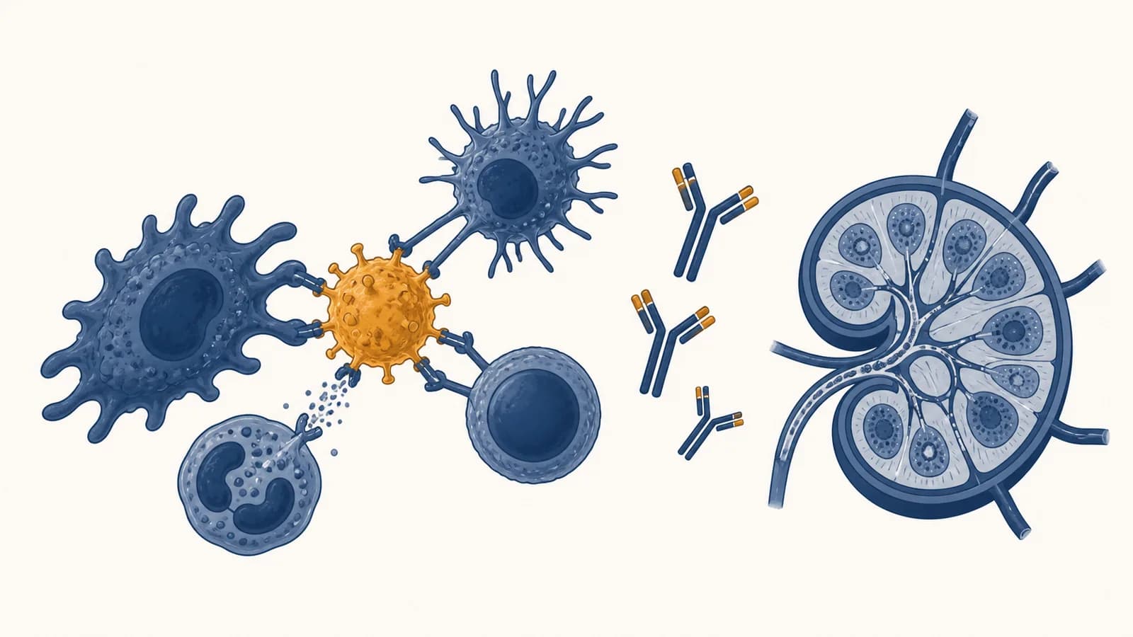
Immunology is a branch of biology that explains how organisms recognize and respond to infection, damaged cells, foreign material, and their own tissues. It studies the immune system, including innate immunity, adaptive immunity, antibodies, antigens, immune cells, vaccination, allergies, and autoimmune disease. Immunity exists because bodies need to remove genuine threats while preserving healthy cells and useful microbes. That task requires recognition, communication, attack, restraint, and memory.
What immunology actually is
Immunology is the study of biological recognition and controlled defense. It asks how cells detect danger, how they coordinate a response, how some responses become faster after exposure, and how failures of recognition produce infection, allergy, autoimmunity, or rejection of a transplant.
The immune system is often described as an army, but the comparison hides its hardest problem. Killing is only one part of the job. The system must decide what to leave alone. A skin cell, a harmless food protein, a gut bacterium, a transplanted kidney, and a virus inside a lung cell present different combinations of location, molecular pattern, and damage. An immune response that attacks everything foreign would injure the body every time a person ate. A response that attacked only obviously damaged tissue would let many viruses reproduce before detection.
Immunologists therefore study decisions made across many scales. Molecules fit together at receptor surfaces. Cells release chemical messages called cytokines. White blood cells move through blood, lymph, and tissues. Organs such as bone marrow, thymus, lymph nodes, and spleen produce, train, or gather immune cells. The clinical effects appear as fever, swelling, recovery, chronic inflammation, or vulnerability to infection.
Recognition is selective, not perfect. Immune receptors respond to molecular features and cellular context. They do not read a label that says “dangerous” or “safe.”
This makes immunology part of the wider study of Biology: it connects cell structure, genetics, evolution, metabolism, and communication to decisions that keep a whole organism alive.
What the immune system is actually made of
The immune system is a distributed network of barriers, molecules, white blood cells, and lymphoid organs. No single organ controls it. Defense emerges as these parts detect changes, exchange signals, travel to tissues, remove targets, and then reduce the response.
Barriers control entry
Skin forms a physical wall of tightly connected cells. Mucus traps particles in airways and the gut. Cilia move airway mucus toward the throat. Tears and saliva wash exposed surfaces and contain antimicrobial molecules. Stomach acid creates a chemical environment that many swallowed microbes cannot tolerate. These defenses act before an invading organism reaches internal tissue.
Barrier defense also involves resident microbes. Organisms already living on skin and mucosal surfaces occupy space and use nutrients, which can make it harder for a pathogen to establish itself. This relationship is studied in the biology of microbes and microbial communities, because the immune system must manage residents as well as invaders.
Cells divide the work
Neutrophils rapidly enter infected tissue and engulf microbes. Macrophages engulf material, release signals, and help repair tissue. Dendritic cells sample their surroundings and can activate T cells. Natural killer cells inspect body cells for signs of infection or abnormal change. B cells can become plasma cells that secrete antibodies. T cells include helper cells that coordinate responses, killer cells that destroy selected infected cells, and regulatory cells that restrain other immune cells.
| Part | Main job | Typical location |
|---|---|---|
| Neutrophil | Rapid engulfment and killing of microbes | Blood, then inflamed tissue |
| Macrophage | Engulfment, signaling, and tissue cleanup | Most tissues |
| Dendritic cell | Antigen sampling and T cell activation | Tissues and lymph nodes |
| B cell | Antibody response and immune memory | Lymphoid tissue and blood |
| T cell | Coordination, cell killing, or restraint | Lymphoid tissue, blood, and tissues |
The names describe common roles, not isolated machines. A macrophage changes its behavior according to the messages it receives. A helper T cell can support B cells or activate other cells. The immune response depends on changing cell states and conversations between cells.
Organs create meeting places
Bone marrow produces blood cells and is where B cells develop. The thymus is where developing T cells are selected. Lymphatic vessels collect fluid from tissues and carry it toward lymph nodes. In a node, antigen from a tissue can meet rare lymphocytes able to recognize it. The spleen performs related surveillance for material carried in blood. Tonsils and other mucosal lymphoid tissues sample exposed surfaces.
A swollen lymph node during an infection is evidence of activity in this network. Fluid, antigen-presenting cells, and lymphocytes are gathering there, and selected lymphocytes are multiplying.
How innate immunity works
Innate immunity responds quickly by recognizing broad signs of microbes, tissue injury, or cellular stress. Its receptors are inherited rather than individually redesigned after infection, so it can act immediately through barriers, inflammation, engulfment, antimicrobial proteins, and the destruction of abnormal cells.
Many innate receptors recognize molecular arrangements that occur often in microbes or appear in the wrong place during damage. A receptor might respond to a form of microbial nucleic acid, a bacterial surface component, or molecules released from broken cells. Detection triggers gene activity and chemical signals. Those signals change nearby blood vessels and recruit more cells.
A cut carries bacteria past the dry outer layers of skin into moist tissue with nutrients.
Macrophages and other local cells recognize microbial features or damaged-cell molecules and release cytokines.
Local vessels widen and become more permeable. Fluid and defensive proteins enter tissue, while vessel walls help white blood cells stop and exit.
Neutrophils and macrophages engulf microbes, release antimicrobial substances, and clear damaged material.
As the trigger disappears, restraining signals limit recruitment and tissue repair becomes dominant.
Redness, heat, swelling, and pain are linked consequences of inflammation. Increased blood flow contributes redness and warmth. Fluid moving into tissue contributes swelling. Chemical mediators and pressure can activate pain-sensing nerves. These effects help deliver defenses and discourage use of an injured area, but an excessive or prolonged response can damage healthy tissue.
Complement marks, recruits, and damages
Complement is a set of blood proteins that activate one another in a cascade. Activation can coat a microbial surface, making the target easier for phagocytes to grab. Small fragments recruit and activate immune cells. In some cases, a terminal complex forms pores in a target membrane. Host cells carry regulatory proteins that normally limit complement damage on their own surfaces.
Innate immunity is not simply a rough opening act. It shapes what comes next. Cytokines and antigen presentation influence which adaptive cells activate, where they travel, and what kind of response they produce.
How adaptive immunity works
Adaptive immunity works by selecting rare B and T lymphocytes whose receptors fit a particular antigen, then multiplying and specializing those cells. The first response takes time, but surviving memory cells can support a faster and often stronger response after later exposure.
Before infection, the body contains many lymphocytes with different receptor shapes. Diversity arises as developing B and T cells rearrange segments of receptor genes. Each cell produces a receptor with a particular binding pattern. The body does not wait for a pathogen and then design a receptor deliberately. Instead, antigen selects from a large existing collection.
Clonal selection turns recognition into numbers
A matching receptor is necessary but often not sufficient for activation. A naive T cell usually needs its receptor to recognize antigen displayed by a major histocompatibility complex molecule, called MHC, plus additional signals from an antigen-presenting cell. These conditions help tie activation to evidence of infection or danger. Once activated, the selected cell divides repeatedly. Its descendants share the same receptor specificity but can adopt different functions.
If one selected lymphocyte completes 10 divisions and every daughter survives, the model gives cells. Real expansion is less tidy because division and cell death vary.
This visible arithmetic explains why selection can work even when the matching cell was initially rare. It also explains the delay. Recognition has to be followed by gene expression, division, specialization, and movement into the affected tissue.
T cells recognize displayed fragments
T cell receptors do not usually bind intact free-floating antigen. Cells cut proteins into peptide fragments and display them on MHC molecules. Killer T cells generally inspect peptides presented by MHC class I, which is found on most nucleated cells. If a cell displays a foreign or abnormal peptide in the right context, a matching killer T cell can trigger that cell to die. Helper T cells generally recognize peptides on MHC class II molecules found on specialized antigen-presenting cells, then provide signals that direct other immune cells.
B cells make soluble recognition molecules
A B cell receptor can bind an intact antigen. With appropriate signals, often including help from a T cell, the B cell multiplies. Some descendants become plasma cells, which manufacture and secrete large quantities of antibody. Others become memory B cells. During some responses, B cell populations undergo mutation and selection that can increase average antibody affinity for the antigen.
The quoted sentence is a summary, not a claim that the system always succeeds. Pathogens mutate, hide inside cells, suppress signals, or attack immune cells themselves. Adaptive responses can also choose a harmful target or fail to shut down.
Antigens versus antibodies
An antigen is a molecular structure that can be recognized by an immune receptor, while an antibody is a protein made by B cell descendants that binds a particular antigenic feature. The antigen is the target; the antibody is one type of targeting molecule.
A recognized structure, such as part of a viral protein, bacterial surface, pollen protein, food protein, or altered body molecule. One object can carry many distinct antigenic sites.
A Y-shaped immunoglobulin protein with binding sites for a particular molecular feature and a stem region that can recruit other immune mechanisms.
Binding is based on shape, charge, and other chemical interactions across contacting surfaces. It is selective, but “one antibody for exactly one organism” is too simple. An antibody recognizes an epitope, a small part of an antigen. Similar epitopes on different molecules can sometimes produce cross-reactivity.
Binding does not always destroy the target
Antibodies can block a virus from attaching to a cell, a process called neutralization. They can coat a target so phagocytes bind it more readily, called opsonization. Their stem regions can activate complement or recruit immune cells. The outcome depends on antibody class, binding location, amount, and the other mechanisms present.
Several antibody classes have distinct distributions and jobs. IgM is commonly produced early in a first response and forms a structure effective at activating complement. IgG is abundant in blood and tissues and supports neutralization, opsonization, and other functions. IgA is prominent in mucosal secretions. IgE participates in defense against some parasites and in immediate allergic reactions. These are functional tendencies, not a ranking from weak to strong.
A positive antibody test does not automatically mean active illness. It may reflect an earlier infection, vaccination, cross-reactivity, or antibodies that have not yet disappeared. Interpretation depends on the test, timing, symptoms, and prevalence of the condition.
Antibodies are useful laboratory tools because their binding can reveal or capture a molecule. Scientists attach detectable tags to antibodies to locate proteins in cells, identify cell types, or measure analytes in a sample. Related methods are central to laboratory tools that use cells and biomolecules.
How immune memory shows up in vaccination
Vaccination presents the immune system with a controlled form or component of a pathogen, or instructions for making an antigen, so adaptive cells can practice recognition without the usual risk of the disease. Memory cells and antibodies can then shorten a later response.
A vaccine does not create a force field at the skin. Its protection begins with the same recognition machinery used during infection. Vaccine antigen reaches antigen-presenting cells. Innate signals, sometimes strengthened by an adjuvant, support activation. Selected T and B cells expand. Plasma cells make antibodies, and a fraction of activated cells persist as memory populations.
On later exposure, pre-existing antibody may block infection early. Memory B and T cells can also react with fewer delays than naive cells. Protection is not identical for every vaccine or pathogen. A rapidly changing surface antigen may escape some existing antibodies. Immunity at a mucosal surface may differ from immunity measured in blood. Immune memory can reduce severe disease even when it does not prevent every infection.
Boosters repeat information after the response contracts
After an immune response peaks, most expanded cells die as signals fade. This contraction prevents the immune system from remaining permanently enlarged. Some antibody-producing cells and memory cells remain, but their numbers and activity can change over time. A booster presents antigen again, expands selected cells, and can improve the quality or amount of the response.
A school asks for vaccination records after a case of a contagious disease. Public health workers are not treating every student as infected. They are estimating who is likely to have protective memory, who needs follow-up, and where transmission could continue.
Vaccination also has a population effect when it reduces the number of susceptible hosts or shortens infectiousness. The size of that effect depends on the pathogen, vaccine, immune response, contact pattern, and uptake. There is no single immunity percentage that applies to every disease.
How immunology shows up in diagnosis and treatment
Immunology appears in clinics through tests that detect antigens, antibodies, immune cells, or inflammation, and through treatments that strengthen, replace, redirect, or suppress immune functions. The same mechanism can help in one condition and cause harm in another, so context controls the goal.
Tests answer different biological questions
An antigen test looks for part of a pathogen or another target in the sample. An antibody test asks whether binding antibodies are present, which may indicate prior exposure or vaccination. A complete blood count measures categories of blood cells but does not identify every immune state. Flow cytometry uses tagged antibodies and lasers to classify individual cells by their surface or internal molecules. Tissue staining can show which cells and proteins occupy a biopsy.
Every diagnostic test has a sensitivity and specificity under defined conditions. Sensitivity is the proportion of people with the condition who test positive. Specificity is the proportion without the condition who test negative. Those properties do not by themselves tell a patient the chance that a positive result is correct, because that probability also depends on how common the condition is in the tested group.
Suppose 100 of 1,000 people have a condition. A 90% sensitive test finds 90 of them. If specificity is 95%, 5% of the 900 unaffected people, or 45, test positive. The positive predictive value is .
The calculation shows why screening a low-risk population can generate surprising false positives even with a good test. It does not mean the test is useless. It means the result must be read alongside symptoms, exposure, timing, confirmatory methods, and the consequences of missing a case.
Treatment can push the response in either direction
Vaccines and some cytokine treatments stimulate selected immune functions. Monoclonal antibodies can block a signaling molecule, tag a cell for removal, prevent a virus from entering cells, or deliver a drug to a chosen target. In cancer immunotherapy, checkpoint inhibitors remove molecular brakes that tumors may exploit, allowing T cells to attack more strongly. The benefit can come with inflammation in healthy organs because the brakes also protect normal tissue.
Transplant medicine often needs the opposite move. A donated organ displays unfamiliar MHC molecules and other antigens, so recipient T cells can treat it as a threat. Immunosuppressive drugs reduce rejection but also reduce some protection against infection and abnormal cells. Matching, monitoring drug levels, and checking organ function are ways clinicians manage this tradeoff.
These treatments make sense alongside the study of organs and body systems, because immune signaling can alter lungs, skin, joints, intestines, nerves, kidneys, and blood rather than staying in an isolated immune compartment.
Four mistakes people make with immunity
Common mistakes treat immunity as a simple measure of strength, assume every symptom comes directly from a microbe, or interpret one test without timing and context. Correcting these errors requires separating recognition, response, tissue damage, memory, and laboratory measurement.
1. Calling every strong response a good response
A useful response is proportionate and directed. Severe inflammation can damage lung tissue, disturb circulation, or injure an organ even while it attacks a pathogen. Allergy is an immune response to an ordinarily harmless trigger. Autoimmunity directs responses toward the body's own molecules. “Boosting the immune system” is therefore an incomplete goal. A person may need a more specific response, a weaker response, or restored regulation.
2. Treating fever as the pathogen itself
Fever is a regulated rise in body temperature driven by host signaling, not simply heat released by microbes. Immune mediators cause the brain's temperature control system to adopt a higher set point. Chills can occur while the body generates and conserves heat to reach it. Symptoms such as aching and fatigue also reflect host signals. This distinction matters because infection and the response to infection both contribute to illness.
3. Assuming “natural” means harmless or permanent
Infection can produce immune memory, but it can also cause tissue injury, long-term complications, transmission to others, or death. Memory after infection varies with the pathogen and person. Vaccination aims to generate useful recognition with less risk than the disease. Neither route guarantees lifelong protection, and neither can be judged by the word “natural.”
4. Reading a laboratory result without its clock
Antigen, pathogen nucleic acid, IgM, IgG, and symptoms can appear and disappear on different schedules. A sample taken too early may lack detectable antibody. Later, antigen may be gone while antibodies remain. Treatments and immune deficiencies can change the schedule. The specimen type and collection quality matter too. A result is a measurement at a time, not the whole history of an infection.
One number tells how “strong” a person's entire immune system is.
Barrier function, cell counts, receptor diversity, antibody levels, signaling, memory, and regulation can differ independently and must be tested for a specific question.
This is why a clinician asks what was measured, why it was ordered, when the sample was collected, and what else was happening. The useful interpretation is narrower than a slogan and more informative.
How allergies happen
Allergy is an immune response to a usually harmless substance, such as pollen, food protein, or animal material. In immediate allergy, IgE antibodies arm mast cells, and later exposure can trigger rapid mediator release that causes itching, swelling, mucus, wheezing, or systemic anaphylaxis.
During sensitization, a susceptible person's immune system makes IgE against an allergen. IgE binds high-affinity receptors on mast cells in tissues and basophils in blood. On later exposure, allergen can connect neighboring IgE molecules and cluster their receptors. The cell then releases histamine and other mediators. Blood vessels become leaky, smooth muscle can contract, mucus secretion can increase, and nerves can produce itching.
Anaphylaxis is a medical emergency. Rapid trouble breathing, throat or tongue swelling, faintness, or symptoms affecting several body systems after an exposure require emergency action and use of prescribed epinephrine according to the person's care plan.
Not every adverse reaction to food is an allergy. Lactose intolerance, for example, results from insufficient digestion of lactose and does not require IgE. Irritation, toxic effects, and drug side effects can also mimic parts of an allergic reaction. Diagnosis combines the clinical history with carefully chosen tests because sensitization on a test does not always equal symptomatic allergy.
How autoimmunity and immunodeficiency differ
Autoimmunity is harmful immune recognition or attack directed at the body's own components, while immunodeficiency is inadequate immune protection caused by missing or impaired defenses. One reflects failed tolerance, the other failed defense, although treatment or disease can make both occur in one person.
Developing lymphocytes are screened against self molecules in the bone marrow and thymus. Cells that react strongly can be deleted, edited, or diverted into regulatory roles. Additional controls act in peripheral tissues. They require more than antigen recognition for activation, suppress self-reactive cells, or isolate certain antigens. These layers are called tolerance. They reduce autoimmunity without deleting every cell that could possibly react with self.
Autoimmune disease occurs when genetic susceptibility and environmental events disturb tolerance enough to injure tissue. The target and mechanism differ among diseases. Antibodies may bind receptors or tissue structures. T cells may attack selected cells. Immune complexes may deposit and inflame tissue. Treatment may block a cytokine, deplete a cell population, or suppress broader immune activity.
Immunodeficiency can be primary, arising from an inherited defect, or secondary, resulting from infection, malnutrition, cancer, medication, or another condition. The pattern of infection can hint at the missing mechanism. A defect in antibodies does not create exactly the same vulnerabilities as a defect in T cells, neutrophils, complement, or skin barriers.
Laboratory investigation follows the mechanism: cell counts, antibody quantities, vaccine responses, complement activity, genetic tests, and functional cell assays answer different questions. A normal result in one branch cannot prove that every branch works normally.
How the immune system knows when to stop
The immune system stops through loss of the triggering antigen, disappearance of activating signals, inhibitory receptors, regulatory cells, anti-inflammatory mediators, and programmed death of expanded cells. Resolution is an active biological program, not simply exhaustion after a fight.
During an infection, survival and growth signals keep selected immune cells active. When microbes and damaged material are cleared, those signals decline. Many effector cells undergo apoptosis, a controlled form of cell death that lets material be removed with limited inflammation. Macrophages clear dead cells and can shift toward repair. Regulatory T cells and inhibitory receptor pathways reduce further activation.
Repair is not the same as returning every tissue to its previous state. Epithelial cells may replace lost cells, but deep injury can leave collagen-rich scar tissue. Long-lasting antigen or failed regulation can sustain chronic inflammation. In that state, attempts at defense and repair occur together, potentially changing tissue structure and function.
A small cut is initially red and tender, then becomes less swollen as debris is cleared and the barrier closes. The fading reaction is evidence that pro-inflammatory signals have been replaced by restraint and repair, not evidence that the immune system has vanished.
Sleep, nutrition, hormones, age, previous exposures, inherited variants, medicines, and other illnesses can change immune performance. None can be reduced to one universal “immunity score.” A useful claim names the branch, measurement, tissue, and condition.
Immunology turns biological detail into testable decisions
Immunology connects molecular binding to whole-body outcomes. Receptor shape affects cell activation; cell activation changes tissue behavior; tissue behavior produces symptoms and recovery. Following that chain makes news about outbreaks, vaccine results, allergies, transplants, cancer therapy, and diagnostic tests easier to judge.
The field also shows how variation and natural selection shape living systems. Pathogens evolve ways to enter hosts or evade recognition. Host populations carry varied immune genes. Within one adaptive response, B cell variants can be selected for improved binding. These processes operate at different scales, but all depend on variation affecting survival or reproduction.
The takeaway: Trace any immune claim through five questions. What is recognized? Which cells or molecules detect it? What action follows? How is the response limited? What measurement would show that the proposed mechanism happened?
Try the questions on an everyday case such as a swollen cut, a vaccine appointment, a seasonal allergy, or a diagnostic result. Replace “the immune system reacted” with the relevant barrier, receptor, cell, signal, tissue, and timing. That move turns a vague explanation into biology that can be checked.